The Most Posterior Aspect of a Typical Vertebra Is the
Human Anatomy and Physiology. Anatomy and Physiology questions and answers.

The Vertebral Column Anatomy And Physiology
The bony structures connected directly to the vertebral body are the.

. The most prominent aspect of the thyroid cartilage corresponds to the vertebral level of. Each pedicle forms one of the lateral sides of the vertebral arch. The arch consists of bilateral pedicles pieces of bone that connect the arch to the body and bilateral lamina bone segments form most of the arch connecting the transverse and spinous processes.
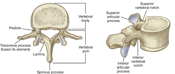
Parts of a Typical Vertebra Part Description Body Central rounded part located anteriorly Pedicle The footpiece attaching to both sides of the body Transverse process Two lateral projections one from each pedicle Spinous process A single projection in a posterior direction Lamina A plate that connects transverse. Are described in Table 82. Kyphosis is defined as an abnormal condition characterized by increased convexity of the thoracic spine curvature the bony structures connected directly to the vertebral body are the pedicles the most posterior aspect of a typical vertebra is the spinous process the joints between articular processes of vertebra are termed zygapophyseal joints.
Spinous process Joints between articular processes of vertebra. The arch consists of bilateral pedicles cylindrical processes of bone that connect the arch to the body and bilateral lamina flat bone segments form most of the arch connecting the transverse and spinous processes. The most posterior aspect of a typical vertebra is the.
T2-3 intervertebral disk space is found. View vertebral quizdocx from HS MISC at Gwinnett Technical College. The only atypical vertebra of the lumbar region is L5.
The pedicles are anchored to the posterior side of the vertebral body. The most posterior aspect of a typical vertebra is the. The most posterior aspect of a typical vertebra is the.
The vertebral arch forms the posterior portion of each vertebra. The vertebral arch forms the posterior portion of each vertebra. L5 also has the most inferiorly located discovertebral unit in the human vertebral column.
Between the superior and inferior articular process. The most posterior portion of a typical vertebra is called the. Bony structures connected directly to the vertebral body.
Pedicles Most posterior aspect of a typical vertebra. The arch along with the posterior aspect of the body forms the vertebral spinal canal which contains the spinal cord. Each pedicle forms one of the lateral sides of the vertebral arch.
The arch along with the posterior aspect of the body forms the vertebral spinal canal which contains the spinal cord. Abnormal increased convexityof the spine. It consists of four parts the right and left pedicles and the right and left laminae.
Spinous process clamina dbody. This online quiz is called superior aspect of typical cervical vertebra. The arch consists of bilateral pedicles pieces of bone that connect the arch to the body and bilateral lamina bone segments form most of the arch connecting the transverse and spinous processes.
The pedicles are anchored to the posterior side of the vertebral body. The joints between articular processes of vertebra are termed. The posterior longitudinal ligament runs within the vertebral canal along the posterior aspect of the vertebral bodies.
Question 1 1 1 pts The most posterior aspect of a typical vertebra is the. At the level of the jugular notch. The vertebral arch forms the posterior portion of each vertebra.
The typical vertebra has a body that points anteriorly Posterior to the body is from BIOS 254 at Biola University. Zygapophyseal joints Where is the articular pillar located on a cervical vertebra. L5 has the largest vertebral body and transverse processes.
What part of a vertebra is most posterior in position. The arch along with the posterior aspect of the body forms the vertebral spinal canal which contains the spinal cord. It consists of four parts the right and left pedicles and the right and left laminae.
The pedicles are anchored to the posterior side of the vertebral body. Each pedicle forms one of the lateral sides of the vertebral arch. It consists of four parts the right and left pedicles and the right and left laminae.
It is attached mainly to the IV discs and less so to the posterior aspects of the vertebral bodies from C2 axis to the sacrum often bridging fat and vessels between the ligament and the bony surface. The anterior aspect of the vertebral body is higher than the posterior aspect contributing to the slightly wedge-shaped appearance it has. The typical vertebra has a body that points anteriorly Posterior to the body is.

Regional Vertebrae Anatomy Anatomy Bones Anatomy Medical Anatomy

Features Of Typical Vertebra Fall 2020 Msu Mediaspace

Human Spine Anatomy Posters Anatomy Bones Human Anatomy Human Spine

Clinically Applied Anatomy Of The Vertebral Column Surgery Oxford International Edition

Vertebral Anatomy Anatomy Bones Medical Anatomy Human Anatomy And Physiology
Back Basicmedical Key

Case 24 Anatomy Flashcards Quizlet

Crossfit Basic Structure Of The Vertebrae

Structure Of A Typical Vertebrae Diagram Quizlet

Atlas C1 Bottom View Human Skeleton Anatomy Human Anatomy Anatomy
![]()
Vertebral Column Anatomy Vertebrae Joints Ligaments Kenhub

The Vertebral Column Anatomy And Physiology

Typical Vertebral Structure Medical Coding Human Anatomy And Physiology Science Revision

Coccyx Posterior Aspect Lower Back Anatomy Anatomy Natural History

Bones Of The Vertebral Column Osmosis

Cervical Vertebra Anterior View Cervical Vertebrae Vertebrae Skull And Bones

Chapter 8 Structure Of Typical Vertebra Flashcards Quizlet
Back Basicmedical Key

Vertebrae An Overview Sciencedirect Topics


Comments
Post a Comment